Nematodes or roundworms themselves (Nematoda) are a type of protostomes, protocavities, bilaterally symmetrical moulting animals.
Spread. Nematodes are one of the most widespread animal species that have been able to colonize a wide range of habitats - from the interstitium (the space between grains of sand) and moss communities to Arctic ice (e.gTheristis Melnikovi and Cryonema crissum, found in the thickness of perennial ice in the central part of the Arctic Ocean).Parasitic nematodes are of particular interest for research, also due to the great diversity of their hosts.
Construction plan. Thin, spindle-shaped body, tapering towards the ends, round cross-section.The mouth is at the front end and the powder (anus) is at the back end.The outside of the body is covered with a multilayer elastic cuticle - a non-cellular formation secreted by the hypodermis.The hypodermis or epidermis is located under the cuticle.The muscles are represented by a layer of longitudinal, obliquely striated muscle fibers.The primary body cavity (schizocoel) without its own epithelial lining is filled with fluid.
Digestive system. The mouth opening at the anterior end of the body is surrounded by projections - lips (usually three) - and leads into a muscular ectodermal pharynx with a triangular lumen.The pharynx leads to the endodermal midgut from a single layer of columnar epithelial cells.Next is a short ectodermal hindgut that opens into the anus.
Excretory system. Excretory organs are unicellular glands that replaced protonephridia.At the front of the body there is usually a neck gland from which a short duct arises.There are also “storage kidneys” - phagocytic organs that accumulate insoluble metabolic products that are not removed from the body.
Circulatory and respiratory systems. These systems are missing.Breathing occurs through the skin.Anaerobic metabolism is also possible (anaerobic breakdown of glycogen to butyric and valeric acid in parasites).
Nervous system. The nervous system is of the squamous type.Represented by a nerve ring and six longitudinal trunks.The two nerve trunks running along the ventral and dorsal lines are stronger and connected by semicircular nerve bridges (commissures).
Sensory organs. There are papillae and setae – organs of touch located around the mouth.Some marine representatives have primitive eyes - pigment spots.Chemical sensory organs, amphibians, usually have the shape of a pocket, a spiral or a slit.They are located on the sides of the headboard and are particularly well developed in males as they help search for females.
Reproduction and development. Nematodes are dioecious animals.The internal sexual organs are paired and have a tubular structure.Reproduction is only sexual.The sexual dimorphism is pronounced: females are larger, in males the rear end of the body is curved.Fertilization occurs internally and viviparity occurs.During development, nematodes go through four larval stages, which are separated by molting and accompanied by the detachment of the cuticle.The third instar in some species (including the famous Caenorhabditis elegans) Under unfavorable conditions it goes into the so-called permanent stage - a resting larva.
Parasitism. Currently, of the more than 24,000 nematode species described, about half are parasitic.They can affect almost all tissues and organs: connective tissue, muscles, blood and lymphatic vessels, gonads, sensory organs as well as the body cavity, etc. These include both ecto- and endoparasites of plants, vertebrates and invertebrates, including other nematodes and even protozoa.
The most important representatives of roundworms from the perspective of medical parasitology are described below.
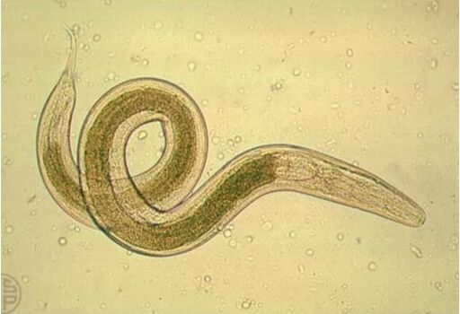
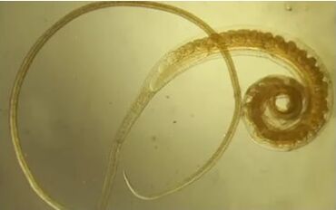
Human roundworm(Ascaris lumbricoides)
Look.The body, which is pointed at the ends, is pinkish-white.Dimensions: Male – 15-25 cm, Female – 20-40 cm.The body is covered with a ten-layer flexible cuticle that protects against mechanical stress and digestive enzymes from the host.
Spread. The species is cosmopolitan - widespread everywhere, but different countries have different percentages of infected people.For example, in Japan, more than 90% of the population is infected with roundworms through the use of human excrement as fertilizer.Roundworms are less common in areas with hot, dry climates.
Life cycle.The development takes place without a change of ownership.Adult worms parasitize the small intestine and cause ascariasis.A person is usually affected by several dozen roundworms (the record is 900 pieces).The lifespan in the intestine is about one year.Like other nematodes, roundworms are dioecious.A sexually mature female lays around 200,000 oval eggs every day, which are released into the external environment in the feces.Roundworms are classified as geohelminths – they require the development of a larval stage in the soil.Under favorable conditions (moist soil with a temperature of around 25 ° C and sufficient access to oxygen), a larva develops in the egg.The development time varies from 16 days to several months and depends on the air temperature.Such eggs containing a larva may be considered invasive.
Infection occurs when eggs are ingested in food or water;Transmission does not occur directly from person to person.In the intestine, the larvae burrow through the intestinal wall, penetrate the blood vessels and liver, and then migrate through the inferior vena cava into the right atrium and right ventricle.From the latter, the larvae migrate through the pulmonary circulation to the lungs, where they pass from the blood into the alveoli, bronchi, trachea and oral cavity.A secondary infection occurs in the oral cavity: the larvae are swallowed, enter the intestines and become sexually mature after three months.The process of “growing up” in nematodes is associated with molting (usually four of them).
Clinical picture of ascariasis. In the migratory stage of ascariasis, cough (helps the larvae enter the throat), chest pain, allergic reactions and fever are observed.
In the intestinal stage, damage to the intestinal mucosa occurs and the body is poisoned with toxic metabolic products.Symptoms: nausea, vomiting, stool disorders, loss of appetite.
Long-term consequences of an infection: general decline in performance, sleep disorders.When worms invade the bile ducts and respiratory tract, the danger is fatal.In addition, roundworm larvae can invade the brain (e.g. from the inferior vena cava to the superior vena cava and then along the brachiocephalic vein) and cause meningoencephalitis, which is associated with migraines.
Prevention. Wash hands before eating and preparing food.Wash vegetables and fruits.Eggs are also carried by flies, so controlling these flies, for example with Velcro, also helps prevent ascariasis.
Interesting fact. There are studies showing the positive effects of roundworm infection on relieving the symptoms of autoimmune diseases and increasing fertility in women.Scientists attribute this to the parasites' effect on the immune system by affecting the amount of T cells in the body.However, the mechanism is currently not well understood to allow reliable conclusions to be drawn.
Pinworm(Enterobius vermicularis)
Look. Gray-white nematode, males 2–5 mm long, females 8–14 mm long.The tail end is pointed (hence the name).A characteristic swelling of the esophagus is noticeable at the front end of the body.
Life cycle.Pinworms parasitize the lower part of the small intestine and colon, causing enterobiasis.The lifespan is 1-2 months.The front end of the pinworm attaches itself to the intestinal wall.A sexually mature female crawls out of the large intestine through the anus and lays 5 to 15,000 eggs on the skin near the anus, after which she dies.
The females crawling out is accompanied by itching.Scratching the skin transfers eggs to the hands and more.Flies are also involved in the transmission of eggs.Infection occurs through ingestion.Larvae hatch from eggs and enter the intestines.
Epidemiology and clinical picture of enterobiasis. Enterobiasis is widespread and is particularly common in children due to non-compliance with personal hygiene rules and overcrowding in kindergartens and schools.Transmission from person to person without an intermediate host.Reduces the effect of vaccinations.
Symptoms: abdominal pain, loss of appetite, headache, allergic manifestations, perianal itching (leads to sleep disorders, increases irritability).
Trichinae(Trichinella spiralis)
Description.Small nematode 2-4 mm long.Parasites the mucous membrane of the small intestine.Distributed in Eurasia and North America.
Life cycle. A change of host is necessary for the development of trichinae.As a rule, these are wild animals (foxes, wolves, bears, wild boars) as well as people and farm animals.The females anchor themselves with the front end of the body in the intestinal epithelium and give birth to 1-2,000 larvae.Ovoviviparity is typical: the larvae hatch from eggs in the female genital tract.The larvae are transported through the body via the blood and lymph vessels and settle in the striated muscles.At this stage they have a stylet with which they destroy muscle tissue, causing the host to form a capsule in which they live curled up in the future.After a few months the capsule is soaked with lime.Such a muscular trichine can exist for several years and survive even after the death of the owner and the decomposition of his corpse.
In the stomach of the new host (after eating the corpse of the previous host), the larvae are released from the capsule, penetrate the mucous membrane and transform into adult worms within a few days after four molts.
Clinical picture of trichinosis. Increased temperature, facial swelling, muscle pain, allergic reactions.
Prevention. Trichinosis is transmitted through food through contaminated meat.Therefore, to prevent the disease, the meat must be subjected to a veterinary examination and properly prepared - boiled for 2-3 hours.Cooking methods such as smoking and salting do not destroy trichinae.
Whipworm(Trichocephalus trichurus)
Look.The worm is whitish in color and is about 4 cm long.The front end is thin and resembles hair (hence the name).
spread.They prefer countries with humid and warm climates.
Life cycle.The worm parasitizes in the initial part of the large intestine, only in humans.Causes trichuriasis.The lifespan of a human being is several years.The thin end penetrates the thickness of the mucous membrane of the intestinal wall.It feeds on tissue fluid and blood.
The female lays 1-3,000 eggs, which are released into the external environment in the feces.Like the roundworm, the whipworm is also related to geohelminths: for the eggs to become invasive, they must remain in the soil for a month at a certain humidity and temperature (25-30 ° C).An infection then occurs when the eggs are swallowed;Larvae hatch from them in the host's intestine, penetrate the intestinal villi and grow there for about a week.After destroying the villi, they enter the intestinal lumen, enter the large intestine, establish themselves there and reach maturity within a month.
Clinical picture of trichocephalosis. The worm damages the mucous membrane of the large intestine and leads to poisoning of the host with waste products.The whipworm is a hematophage and can therefore cause anemia.Trichocephalosis is accompanied by abdominal pain, headaches and dizziness.Because the whipworm attaches itself to the intestinal wall, it is more difficult to remove from the host than other parasites.
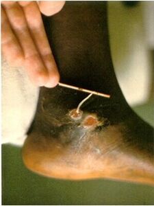
Rishta(Dracunculus medinensis)
Look.A thin, whitish nematode, females 30–120 cm long, males no longer than 4 cm.There is a small stinger on the tail.
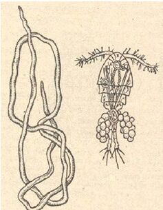
Spread: tropical countries of Asia and Africa.
Life cycle.Infection occurs when drinking unboiled water that contains copepods.The crustaceans in the stomach die under the influence of hydrochloric acid, but the guinea worm larvae survive and are distributed throughout the body through the lymphatic system.They then penetrate the body cavity, shed their skin there and reach sexual maturity.After mating, the male dies and the female migrates to the subcutaneous tissue, where a purulent abscess forms, which is accompanied by burning and pain.Cool water is best for pain relief.
The development of eggs forces the female to move her “head” forward towards the surface of the skin, creating an inflammatory process on her way, which turns into a purulent abscess, which then bursts.When the female's uterus enters the water, it ruptures and the larvae hatching from the eggs come out.To ensure that development is not interrupted, the larvae must infect the cyclops crab, which acts as an intermediate host.The larvae remaining in the water die.After the crustaceans are swallowed by the final host under the influence of stomach acid, the crustaceans dissolve and the larvae easily penetrate the intestine, penetrate through its walls and end up in the lymph nodes, where the cycle of development continues.The disease caused by Guinea worm is called dracunculiasis.
Dracunculiasis.The incubation period lasts up to nine months and ends when the female reaches sexual maturity.And in a person who has already suffered from dracunculiasis, purulent abscesses begin to form at this time.The only salvation from the pain is a pond.Relief is immediate, but upon contact with water the blisters burst and the Guinea worm throws the larvae into the water.The crustaceans eat them and the life cycle begins again.
Treatment for dracunculiasis often involves making an incision at the site of the blister and gradually pulling out the worm and wrapping it around a stick.This takes days, sometimes weeks (you have to pull the worm out slowly and carefully so it doesn't break).It was believed that the appearance of a guinea worm wrapped around a stick became a kind of prototype for the symbol of medicine - the staff of Asclepius entwined with a snake.
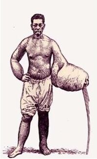
Bancroft's thread (Filaria) or Bancroft's thread(Wuchereria bancrofti)
Look.White thread nematode, female 10 cm long, male 4 cm long.
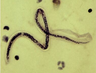
Distribution. Tropics, subtropics of Asia, Africa, Central and South America.
Life cycle. In adults, they usually occur in the lymph glands and vessels, which obstructs the drainage of lymph and leads to persistent swelling.Females produce larvae - nocturnal microfilariae that appear in the peripheral blood at night and penetrate deep into the body during the day (into the pulmonary vessels and kidneys).This is because the intermediate hosts are mosquitoes, which mostly suck blood in the evening and at night.The larvae enter the mosquito's stomach and then into the body cavity, where they grow.They then accumulate near the trunk, from where they are transmitted to humans through bloodsucking.Bancroft filaments cause elephantiasis or elephantiasis or elephantiasis.It should be noted that this disease can also be caused by other nematodes.
Clinical picture and treatment of elephantiasis. Enlargement of any part of the body occurs due to hyperplasia (painful growth) of the skin and subcutaneous tissue caused by inflammatory thickening of the walls of lymphatic vessels and stagnation of lymph caused by blockage of lymphatic vessels by adult Bancroft's threads.The skin on the affected part of the body becomes covered with ulcers.
Treatment for elephantiasis is aimed at improving fluid drainage.The use of anthelmintics is effective.In later stages, surgery may be required.